Canine mast cell tumors: An Overview for Dog Owners
Did you comprehend that Mast Cell Tumors in Dog are among them? Therefore the most common types of tumors on the skin among dogs? You’ll learn more about canine mast cell tumors. In this educational essay. Discussing their causes, signs, and solutions.
Granules contain bioactive chemicals like heparin. And histamine is abundant in mast cells. When they encounter an allergen or parasite. For example, they can break down, or release. Although their granule contents into the environment. The granules’ compounds produce swelling. Redness, and irritation when this occurs.
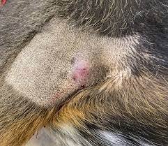
It could be that you saw a small bump on your dog which appears to form overnight. Or it could have been years since the mass appeared. However, it becomes red and swollen all of a sudden. And your dog won’t stop licking it. Alternatively, your dog might have experienced a lump. That you were not aware. Of until the veterinarian noticed them. During a routine check-up.
A mast cell tumor could be the concern. Diagnosis in each of these cases. For canines with this most common of skin tumors. A mast cell tumor identification opens up. A whole new set of issues. Malignant mast cells can proliferate in one part of the body. And result in a mast cell tumor. Whereas healthy mast cells are robust and resistant. To disintegration, tumor cells are delicate.
What exactly is a canine Mast Cell Tumors in Dog?
Mast cells are part of the immunological system’s apparatus. Include white blood cells. Their purpose is to fight foreign invaders. Which are parasites but can also be allergies like dust or pollen.
Mast Cell Tumors in Dog contain large amounts of granules that contain active compounds. Including histamine and heparin. They can disintegrate or leak their particle. Contents into the surroundings. In response to allergens or parasites, for instance. As a result in swelling, redness, and inflammation. Due to the substances in the granules.
A mast cell tumor can develop. When malignant mast cells multiply in one area of the body. The tumor cells are brittle. And prone to degranulation, in contrast to healthy mast cells.
Mast Cell Tumors in Dog can cause modest symptoms like discomfort. And swelling in the vicinity of the tumor when they degrade. The dog may lick or scratch the mast cell tumor as a result of this. In particular, the region in question may expand, and turn red. And then return to its normal size when the mass touches and presses.
More serious systemic symptoms. Such as gastrointestinal ulcers or bleeding. And bruises in the surrounding tissues could happen to the dog. Dogs impacts may also experience a drop in blood pressure. Or may pass away from an asthma attack, or a severe allergic reaction.
What Leads to Mast Cell Tumors in Dog?
The cause of mast cell tumors is unknown. Like that of the majority of tumors. They have links to the use of irritating chemicals. On the skin or persistent inflammation. Research has shown that Boxer dogs with MCTs have higher levels. Of chromosomal fragile site expression. Which is an unstable genomic locus. Prone to breakage or rearrangement.
This condition is likely to condition humans. To develop instances of certain malignancies. This research focuses on young boxers who were not pregnant. Therefore believe that the age difference was the cause. Of the enhanced expression. It is unclear exactly which genetic changes. In dogs raise the risk of cancer in people.
In certain dogs, alterations in the p53 tumor-suppressing. Pathways have been seen. It demonstrates that several dogs. Have an aberrant synthesis of proteins. Including p21 and p27, cyclin-dependent kinase. (an enzyme stopper that inhibits. The activity of one or more protein kinases). And cell cycle inhibitors.
Canine MCTs have recently been shown to contain c-Kit. A tyrosine kinase receptor is necessary. For the proliferation of hematopoietic stem cells. Which are multifaceted stem cells that give rise to all forms of blood cells. In addition, several investigations have hypothesized abnormalities. In the c-Kit juxtamembrane region.
Which is next to a transmembrane on one side of it. This could result in constitutive activation. Or the steady synthesis of an enzyme or protein. In the absence of hematological growth stem cell factor. Studies suggest that the hormones progesterone and estrogen. May also affect mast cell cancers, albeit this is not understood.
What Signs Do Mast Cell Tumors Present With?
There is a wide range in the clinical symptoms of mast cell tumors.
- While 11–14% of tumors include numerous infections. The majority of tumors are solitary.
- 1-4 cm in diameter, well-different tumors are solitary.
- They have a rubbery look and grow.
- Usually, they last for at least six months.
- Although they do not cause ulcers, the skin may lose its hair.
- Ulcerating, heterogeneous mast cell tumors. Can be quite uncomfortable.
- Mast cell tumors enlarge more than other types of tumors.
- Little satellite nodules may form in the surrounding tissues. And the tissues surrounding them. May become fluid-filled and inflammatory. Tumors classified as intermediate lie. In the middle of these two extremes.
- The symptoms of vomiting, an eating disorder. And melena black, tarry stools. Linked to gastrointestinal bleeding may coexist with stomach ulcers.
Visceral MCT is rare, although it can happen to dogs. Usually, a primary cutaneous lesion. That is not distinguished comes before it. Examples of anomalies that manifest. Includes (enlarged liver), (expanded spleen). And lymphadenopathy (enlarged lymph nodes). Carcinogenic mast cell tumors. Often entail the involvement of the bone marrow. And circulation in the peripheral regions. There have been reports of neoplastic cell-containing. Pleural and peritoneal effusions. Or surplus fluid in the abdominal and pleural cavity.
What are the Mast Cell Tumor Therapies?
When diagnosing mast cell tumors, aspiration with a fine needle. And immunohistochemistry (FNAC) is the initial step.
Occurring MCTs (mostly undifferentiated MCTs). Require alternative diagnostic techniques. Such as histology, specific stains, etc. If the MCT is in a place. Where wide surgical excision is feasible. This can be determined from the diagnosis. If that’s the case, surgery finishes right away. The removed tissue is provided. For histologic grade evaluation.
For grading, histologic assessment top choice as FNAC is insufficient. A second diagnosis occurs only. If the lesion turns out to be homogenous. With incomplete surgical margins. Further diagnostic testing occurs to stage the sickness. If the lesion’s location happens. To be unsuitable for surgery.
A thorough blood test and a buff-colored coat smear. To determine peripheral mastocytosis. Thoracic radiography, bone marrow aspiration. Abdominal ultrasonography. With a cytologic examination of the spleen or liver. An exhaustive blood count is all part of comprehensive staging. An incisional biopsy serves to determine the histologic grade.
Abdominal ultrasound, and needle. Aspiration cytology, for the localized node. Routine geriatric pre-anesthetic blood tests are the steps. That makes up pre-surgical staging. A chest radiograph is insufficient to diagnose tumors. On the other hand, they help rule out unrelated illnesses. Or concealed heart problems that can complicate anesthesia.
An infrequent single lymph node cell is not concerning. In terms of lymph node cytology. If they arrive in bunches, though, that should raise some red flags. Inhaling bone marrow, smears of the buffy coat. And aspirating regularly. Functioning abdominal organs is not advised.
In these cases, it is recommended. To perform surgery on a cytologically. Suspect node so that a histological examination can run. Before surgery, digital palpation serves. To determine the extent of metastases. It is higher by using ultrasound. Or computerized tomography for diagnosis.
Which Two Mast Cell Tumor Treatments Are the Most Effective?
Currently available, are the two most successful treatments. Techniques are radiation therapy and surgical excision. Since tumors stay small and amenable to a broad excision. Surgery is the recommended method of treatment. Antihistamine medication should utilize the excision. For MCTs, surgery with a 3 cm margin. Of neighboring normal tissues has been recommended.
According to a more recent study, most MCTs can be completely. Removed with 2 cm lateral margins. Deep margins are equally important as lateral profit margins. And it is possible to remove one fascial plane. That is deep into the lesion.
If essential, deep-lying muscle layers inside the tumor are cut. Every surgical margin should have a histological examination done. An incisional sampling should be used to determine. The histologic grade of cancers that are not curable.
Are mast cell cancers similar?
No. Like other cancers in human and veterinary medicine. Some tumors provide a greater risk than others. Some tumors cannot be cut surgically due to their location. A biopsy or complete surgical excision of the mass is necessary. To determine the tumors that are more to be larger and more serious.
After reviewing the biopsy sample, a board-certified veterinary scientist diagnoses it. The type of cancer and assigns a descriptive grade. That indicates the course of the disease and the possibility of recovery. The two most widely utilized grading schemes are the new. Two-tier method and the three-grade system.
We employ the new two-tier grading system (high or low). At the University of Michigan, Veterinary Diagnostic Laboratories to more accurately forecast prognosis. The main method of identifying whether a tumor is a low-grade tumor. (less aggressive, less likely to have detrimental long-term consequences for one’s health).
More likely to be cured by removal with surgery alone) or high-grade (more likely to be possessive. With a greater likelihood for immortality. May benefit from different therapy) is too close. Examine different characteristics of the cancer cells.
Your veterinarian may offer more specialist testing. Such as a mast cell tumor predicting panel, based on the grade to assist in assessment. Whether and what kind of supplemental therapy may be beneficial for your pet.
What Other Mast Cell Tumor Treatment Options Are There?
Hyperthermia is sometimes known as cardiac arrest or sunstroke. Is one of the alternative treatment options. To create the condition in question, drugs. Like photodynamic therapy, an intralesional steroid medication, and cryotherapy. (which involves applying low temperatures locally or to treat medical conditions.
Local surgery or radiation therapy applies only to these lesions. If thorough staging fails to detect spread. The result is not very pleasing because of the localized and distant metastases. Given that high-grade cancers have a guarded prognosis. A coarse fraction radiation treatment strategy.
Adjuvant chemotherapy is beneficial in the great majority of instances. According to recent studies. Vinblastine (2 mg/m2 intravenously) is used in this scenario. For 4 treatments per week, then 4 more treatments are given every 2 weeks. When using VBL, a medicine with steroids. Such as prednisone is first administered orally at a dose of 2 mg/kg for one week. Then 1 mg/kg every other day for two weeks, and finally 1 mg/kg every other day. After the VBL treatment is finished, prednisone is withdrawn.
Sometimes ancillary therapy is recommended for MCT systemic symptoms linked to degranulation. Giving the H1 blocker diphenhydramine (2 to 4 mg/kg given by mouth two times daily). And the H2 blockers a medication called (4 mg/kg given roughly threefold daily). Or ranitidine (2 mg/kg given once daily) can inhibit the effects of histamine production.
Another medication is known as a medication called (0.5 to 1 mg/kg taken orally once a day). Has shown promise in managing the clinical signs. And symptoms of gastrointestinal ulcers linked to gastrinoma. It suggests taking oral sucralfate (0.5 to 1 g) three times a day.
Are there any additional tests or procedures needed?
According to the tumor grade, the pathologist may advise. Doing an MCT diagnostic panel or parts of this panel. Which tumors are more likely to behave benignly? And which are more likely to behave aggressively, demanding extra therapy. This can be further predicted using the panel’s data. Since this panel was born using the original biopsy material. Your pet does not require any further procedures.
Tyrosine kinase inhibitors, or TKIs, are the term for these medications. In veterinary medicine, this is the first instance of “targeted drug management.” For dogs with this mutation and severe mast cell cancers, there is therefore new hope.
The dimension of the tumor and the probability. That it has metastasized or spread to different organs. Or tissues (such as lymph nodes) are additional crucial criteria in establishing. The most successful treatment approach considering. The skin is an organism with multiple layers. The majority of the time and surgical removal is performed.
However, depending on length and location. Your veterinarian could submit the tumor to a lab. Like ours for margin assessment following surgical tumor excision. To provide veterinarians with the most precise information regarding the question and whether or not a mast cell tumor has expanded to the surgical margins.
Your veterinarian may also recommend performing additional tests to ascertain. Whether the tumor has spread to other organs, often the liver and spleen. Or to the lymph glands closest to the tumor site, also known as “regional lymph nodes”. If there is evidence of systemic contribution—a sign that the dog’s. Internal systems are affected—more chemotherapy may be recommended.
Which stages do mast cell tumors go through?
The mast cell tumor stage is crucial. Because it provides insight into the tumors’ internal organ distribution. A tumor is staged following surgical removal and examination of surrounding lymphatic systems. Therefore the number of tumors existing and the involvement of lymph nodes. Are the elements that determine the stage.
0 Stage: One skin tumor that has been partially excised, with no involvement of lymph nodes.
1 Stage: One skin tumor without involvement of lymph nodes.
2 Stage: A single cutaneous tumor including lymph nodes
3 Stage: Several massive, deep cutaneous tumors that may or may not include lymph nodes.
4 Stage: Several tumors that have spread to the skin and impacted lymph nodes. Substages a and b of this stage are further separated. Into those with no additional symptoms and those with additional clinical symptoms.
Conclusion
In conclusion, to give their dogs quick and proper care. Dog owners must have a thorough awareness of mast cell tumors in dogs. Therefore this page discusses mast cell tumors in several ways. Including their etiology, symptoms, and available treatments. Mast cell tumors may occur with a wide range of clinical signs and from small. Slowly growing lumps to more aggressive and uncomfortable tumors.
The study offered valuable perspectives on post-operative management. And observation, emphasizing the necessity of continuous assessment to identify. Any recurrences or systemic involvement. The staging of mast cell tumors focuses on the quantity and location. And involvement of lymph nodes is essential to understanding. The overall prognosis and planning future therapeutic approaches.
In summary, dog owners want to work closely with their physicians. To manage the intricacy of mast cell tumors. To give their furry friends the best care and outcomes possible.
FAQs
What are canine mast cell tumors?
Mast cell tumors are among some of the most common forms of body tumors in canines. These tumors originate in the mast cells. Which are white body blood cells and are embroiled in the immune system’s response. To defend against foreign attackers.
What signs and symptoms can dogs have of mast cell tumors?
Also, Although there are many different symptoms, including lumps. Or tumors on the skin. Abrupt redness and expansion, and behavioral changes. Excessive licking is a typical indicator. Therefore In addition, Systemic symptoms such as nausea. And digestive disorders can appear in more serious cases.
Are canine mast cell cancers all the same?
No, they differ in intensity. A portion of the opposition assesses using biopsies and grading systems. Although The two-tier grading system (good or poor) to evaluate. The prognosis and prospects for recovery.
What part does staging play in malignant Mast Cell Tumors in Dog?
To comprehend how the cancerous cells have spread, staging is essential. It entails assessing the quantity, and size. And the involvement of lymph nodes in the malignancies. Therefor tumors in Stage I are isolated, while Stage 4 tumors have spread throughout the body.
Do dogs with mast cell tumors have other choices for medical care?
Alternative therapies are being investigated, including tyrosine kinase inhibitor-targeted medication management, radiation therapy, and heat and their effectiveness is still being investigated, though.
Canines suffering from advanced mast cell malignancies have hope?
For dogs with particular genetic mutations and severe mast cell malignancies, yes, focused medication therapy with tyrosine kinase inhibitors such toceranib protease (Palladia) and then masitinib mesylate (Kinavet-CA1) gives new hope. Therefore these drugs are an innovative technique in veterinary care.